Categories
Change Password!
Reset Password!


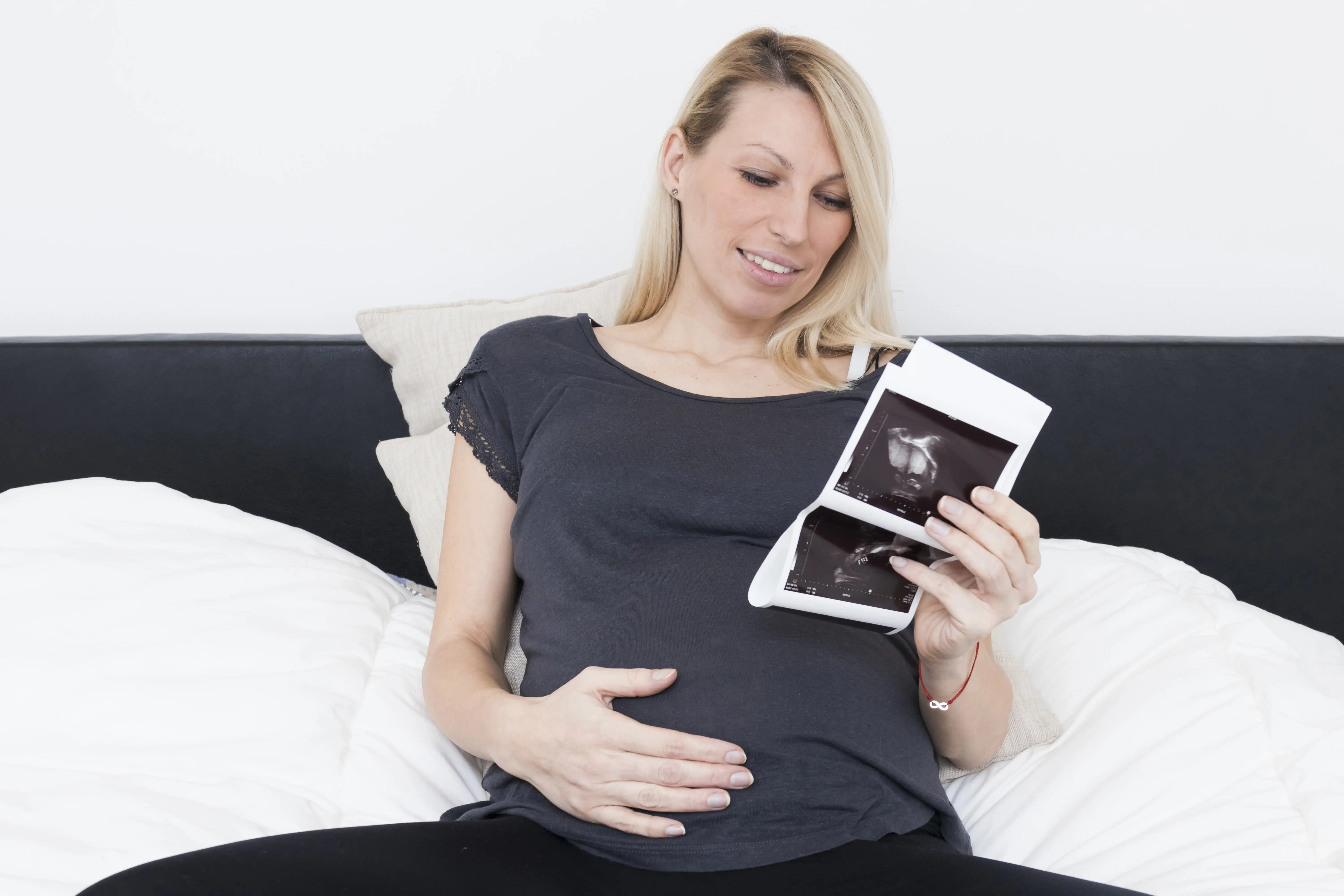
A 22-year-old multigravida Somali woman at 10 weeks’ gestation presented with worsening abdominal pain and was found to have a viable intrauterine pregnancy alongside a ruptured right tubal ectopic pregnancy with hemoperitoneum. She underwent successful surgical management with preservation of the intrauterine pregnancy, which progressed to term with delivery of a healthy neonate.
This case highlights that heterotopic pregnancy can occur naturally and suggests female genital mutilation may be a potential risk factor. Clinicians should maintain a high index of suspicion for heterotopic pregnancy when evaluating pregnant women with acute abdominal pain, and early adnexal ultrasound assessment is essential for timely diagnosis and optimal outcomes.
A 22-year-old Somali woman, gravida 2 para 1, at 10 weeks’ gestation, presented with a 1-week history of progressively worsening lower abdominal pain accompanied by vomiting. The pain was initially cramp-like and confined to the suprapubic region, but later escalated in severity and spread to involve the entire abdomen.
Introduction
Heterotopic pregnancy, a rare obstetric phenomenon, involves the simultaneous implantation of 2 or more embryos at different sites within the reproductive system. Its occurrence varies from about 1 in 30,000 in pregnancies from spontaneous conception to as high as 1 in 100 following assisted reproductive techniques like in vitro fertilization (IVF). Typically, it presents as a concurrent intrauterine pregnancy alongside an extrauterine (ectopic) gestation. The fallopian tube remains the most common site for ectopic implantation in both spontaneous and assisted cases, followed by the cornual region.
Incidences of heterotopic pregnancies in less common locations like the cervix, ovary, or abdominal cavity are exceedingly rare. It shares similar clinical features and risk factors with ectopic pregnancy. Factors that amplify the risk encompass older maternal age, early sexual debut, multiple sexual partners, pelvic infections, a history of infertility, usage of fertility treatments, previous ectopic pregnancies, and prior pelvic surgeries such as female genital mutilation. The likelihood of detecting heterotopic pregnancy is highest, about 70%, between 5 and 8 weeks of gestation.
However, it falls considerably to nearly 10% after 11 weeks. Additionally, the presence of a healthy intrauterine pregnancy during ultrasound examination can complicate the identification of the coexisting ectopic pregnancy, sometimes triggering missed or delayed diagnosis. Although the occurrence of ectopic pregnancy continues to rise, mortality rates have dropped in developed countries, largely because earlier diagnosis prevents tubal rupture, which happens in more than 70% of cases.
In contrast, diagnosing heterotopic pregnancy remains cumbersome in developing countries, often resulting in delayed detection and presentation as a ruptured ectopic pregnancy. This delay is frequently due to limited access to high-resolution ultrasound technology, leading to a higher rate of misdiagnosis of heterotopic pregnancies in these settings. Delayed diagnosis of heterotopic pregnancy can trigger a higher likelihood of complications and death for both the mother and the intrauterine fetus. Despite these risks, nearly 70% of cases elicit a favorable outcome for the intrauterine pregnancy, comparable to that seen in singleton pregnancies.
Medical History
The patient had undergone female genital mutilation at 7 years of age. She had no known personal or family history of congenital abnormalities or chronic illnesses, including diabetes mellitus, hypertension, cardiac or renal diseases. Furthermore, she denied any previous pelvic infections or sexually transmitted infections.
Discussion
Heterotopic pregnancy, the coexistence of intrauterine and extrauterine pregnancies, is extremely rare in natural conception. However, it is considerably more common in pregnancies resulting from assisted reproductive technologies, with incidence rates reaching up to 5% after IVF. In this case, the woman conceived naturally, and the ectopic pregnancy was located in the fallopian tube, the most common site for heterotopic pregnancy. Detection of heterotopic pregnancy becomes progressively challenging as gestation progresses because the enlarging uterus can obscure the extrauterine pregnancy.
Apart from female genital mutilation, which may heighten the risk of pelvic infections and ectopic gestation, no other significant risk factors were present. Heterotopic pregnancy usually manifests symptomatically during the first trimester, typically between 5 and 10 weeks, with clinical signs similar to ectopic pregnancy; however, it can remain asymptomatic in later trimesters. This patient presented at 10 weeks of gestation with abdominal pain, vomiting, tenderness, and suprapubic fullness but without signs of shock. Diagnosis was made using transabdominal Doppler ultrasound, which revealed elevated blood flow in the adnexal mass, though transvaginal Doppler ultrasound is the preferred diagnostic tool due to its higher sensitivity and specificity.
Given the acute presentation with hemoperitoneum, prompt surgical intervention via laparotomy was executed, successfully removing the ectopic pregnancy while preserving the intrauterine gestation. Timely diagnosis is critical as delays magnify maternal and fetal morbidity and mortality; fortunately, about 70% of intrauterine pregnancies in heterotopic pregnancy cases result in favorable outcomes when diagnosed before rupture. In this case, despite late diagnosis, the patient delivered a healthy female neonate weighing 3000 grams with Apgar scores of 8 and 9 at 1 and 5 minutes, respectively, demonstrating that conservative management following surgery can lead to positive maternal and fetal outcomes.
Learning
Heterotopic pregnancy, though rare in natural conception, can occur even without typical risk factors, and female genital mutilation may be a potential contributor. An intrauterine pregnancy on imaging does not eliminate the chances of a concurrent ectopic pregnancy. Early first-trimester adnexal ultrasound is critical for timely diagnosis, especially in women with acute abdominal pain and an adnexal mass. Prompt surgical management of the ectopic component can preserve the intrauterine pregnancy and lead to favorable outcomes.
Journal of Medical Case Reports
Ruptured heterotopic pregnancy with successful term intrauterine pregnancy outcome after natural conception: a case report
Hassen Mohammed Areys et al.


Comments (0)