EN | UA
EN | UA

Help Support

Back

Axial Gout Presenting as Chronic Lumbar Pain in a Young Adult

Chronic lumbar pain, Facet joint gouty arthropathy Chronic lumbar pain, Facet joint gouty arthropathy

See All

Poster abstract

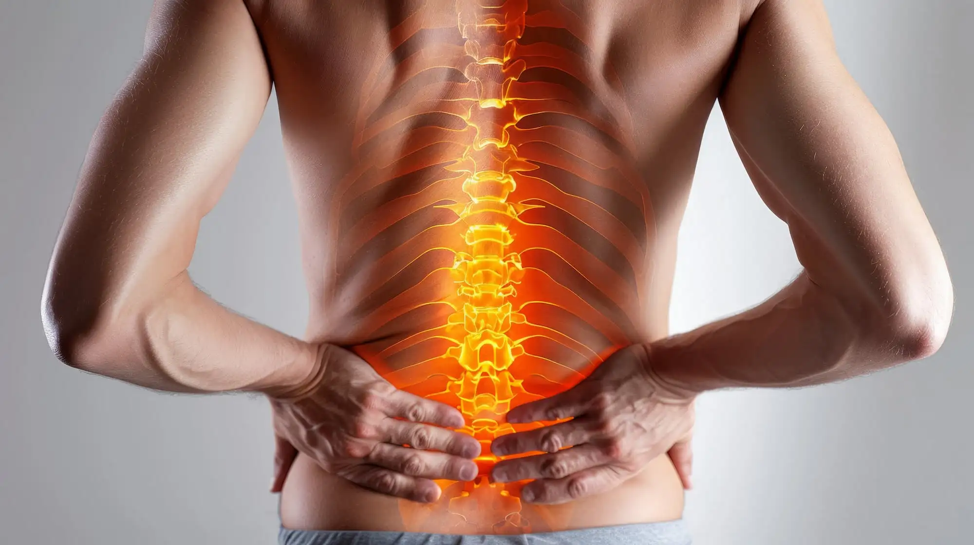
Gout is a common metabolic disorder affecting peripheral joints; however, axial involvement remains rare and often underdiagnosed. A 27-year-old obese male with a ≥10-year history of poorly controlled gout presented with persistent left-sided lower back pain. MRI revealed T1-weighted enhancement and punched-out erosion at the L4-L5 facet joint, consistent with facet joint gouty arthropathy.

This case emphasizes the importance of considering spinal gout as a differential diagnosis in those with chronic back pain and longstanding hyperuricemia. Early recognition is critical, as tophaceous deposits can cause nerve compression, instability, and chronic pain. This case underscores the need for clinician awareness of atypical gout presentations to prevent irreversible spinal complications and boost functional outcomes.

Complaints

  • A 27-year-old man presented with chronic left-sided lower back pain radiating to the left thigh, persisting for 6 months.
  • The patient reported intermittent lower back discomfort over the past 3 years, which had gradually increased in frequency and intensity.
  • Pain was aggravated by spinal flexion and extension, as well as by prolonged sitting, standing, or lying down, leading to significant interference with daily activities.
  • Despite persistent symptoms, the patient denied any paraesthesia, numbness, or weakness in the lower limbs.

Anamnesis

Introduction

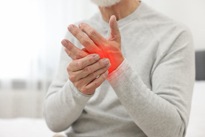
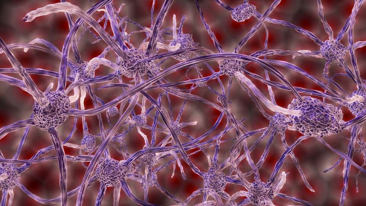
Gout is a metabolic disease marked by monosodium urate crystals accumulation in soft tissues and joints, leading to inflammation and structural impairment. While peripheral joints are most commonly affected, axial involvement is increasingly recognized in patients with chronic or poorly controlled disease. Lumbar facet joint gout may present with chronic back pain, radiculopathy, and functional impairment, frequently mimicking common spinal pathologies. Early identification is critical to prevent neurological complications and irreversible structural deterioration.

Medical History

  • The patient was diagnosed with gout at 17 years of age, initially presenting with pain and swelling in both feet and ankles, consistent with early-onset gout.
  • He later underwent right ankle arthroscopic debridement and os trigonum excision, where chalky urate deposits were noted intraoperatively. His serum uric acid level measured 707 μmol/L, confirming the diagnosis.
  • He had a history of persistent hyperuricemia but showed poor adherence to allopurinol therapy, resulting in recurrent gout flares and gradual disease progression.
  • The patient was morbidly obese (body mass index [BMI] >40), a factor that likely worsened his metabolic profile and contributed to elevated uric acid levels.
  • Although renal function remained within normal limits, underlying metabolic risk factors such as obesity and dietary imbalance were present and likely played a role in disease persistence.

Examination

  • On physical assessment, there was localized tenderness over the L4–L5 facet joint, accompanied by restricted lumbar mobility in all directions.
  • The straight leg raise test revealed tight hamstrings at 60° bilaterally, but no evidence of sciatica.
  • Neurological assessment of the lower limbs illustrated normal tone, power, sensation, and reflexes, indicating no neurological deficit.
  • Visible gouty tophi were noted over the right elbow and lateral malleolus, suggesting chronic urate deposition.
  • Lumbar X-ray demonstrated mild loss of lumbar lordosis with preserved vertebral heights and disc spaces, showing no signs of structural collapse.
  • Contrast-enhanced magnetic resonance imaging (MRI) of the lumbar spine showed T1-weighted enhancement at the L4–L5 facet joint with punched-out erosion. The findings were found to be consistent with facet joint gouty arthropathy.

Treatment

  • The follow-up uric acid measurement was 594 μmol/L. The patient was restarted on allopurinol, which was gradually titrated up to 400 mg daily under rheumatology supervision to achieve optimal urate control.
  • The acute pain flare was managed with a combination of etoricoxib, a short course of oral prednisolone, and low-dose colchicine for prophylaxis against recurrent attacks.
  • A comprehensive lifestyle modification plan was initiated, including a structured weight management program and dietary counseling focused on reducing purine intake and improving overall metabolic health.
  • The patient was placed under regular multidisciplinary follow-up, involving rheumatology, physiotherapy, and nutrition teams, to monitor symptom progression, uric acid levels, and functional recovery.

Results

  • Following initiation of urate-lowering therapy (ULT) and lifestyle interventions, the patient showed gradual improvement in back pain and enhanced functional mobility over the following months.
  • Serum uric acid levels declined steadily, correlating with the resolution of symptoms and better quality of life.

Discussion

This case highlights the uncommon occurrence of spinal involvement in gout and emphasizes the importance of early and continuous ULT to prevent atypical disease presentations. Spinal gout must be suspected in patients with chronic gout, obesity, and male gender who present with persistent back pain. While spinal gout is usually reported in older adults, it can occasionally appear in younger individuals with long-standing or poorly controlled disease and additional risk factors like renal dysfunction.

Diagnosis is cumbersome owing to overlapping symptoms with more common spinal disorders such as disc herniation or spinal stenosis. MRI and histopathology remain diagnostic mainstays. But, dual-energy computed tomography (DECT) offers a reliable, non-invasive alternative with high sensitivity and specificity for urate deposits, despite its higher cost and limited early-stage sensitivity. Spinal gout remains underrecognized and underreported, with limited evidence on long-term outcomes and optimal treatment.

Management parallels that of peripheral gout — acute flares are treated with non-steroidal anti-inflammatory drugs (NSAIDs), colchicine, or corticosteroids, while long-term ULT (like allopurinol or febuxostat) prevents recurrence. Addressing modifiable risk factors like obesity, alcohol use, and dietary habits is essential.

A multidisciplinary approach involving sports medicine, rheumatology, and pain specialists is recommended for accurate diagnosis and comprehensive care. From a sports medicine standpoint, this case emphasizes that not all back pain is mechanical; systemic metabolic disorders like gout can underlie musculoskeletal symptoms. Recognizing spinal gout as part of the differential diagnosis in patients with known gouty disease can trigger more targeted interventions and better patient outcomes.

Learning

  • Axial or spinal gout, though rare, should be considered in patients presenting with persistent lower back pain—particularly young males with long-standing or poorly controlled gout and metabolic comorbidities such as obesity.
  • Prompt diagnosis using advanced imaging (MRI, DECT) is essential to prevent irreversible joint damage, neurological complications, and chronic disability.
  • Axial gout can masquerade as degenerative or mechanical spinal disease, delaying appropriate treatment if not included in the differential diagnosis.
  • Optimal care requires ULT, anti-inflammatory control, and lifestyle modification, including weight management and dietary changes.
  • Coordination between rheumatologists, sports medicine specialists, pain physicians, and rehabilitation experts ensures holistic management—addressing both metabolic control and musculoskeletal recovery.
  • Rehabilitation strategies focusing on mobility, strength, and posture correction are key for long-term pain relief and quality of life improvement.

Source:

Cureus

Article:

Facet Joint Gouty Arthropathy: An Uncommon Cause of Chronic Lumbar Pain

Authors:

Cara C. Chua et al.

Comments (0)

You want to delete this comment? Please mention comment Invalid Text Content Text Content cannot me more than 1000 Something Went Wrong Cancel Confirm Confirm Delete Hide Replies View Replies View Replies en
Try: