Categories
Change Password!
Reset Password!


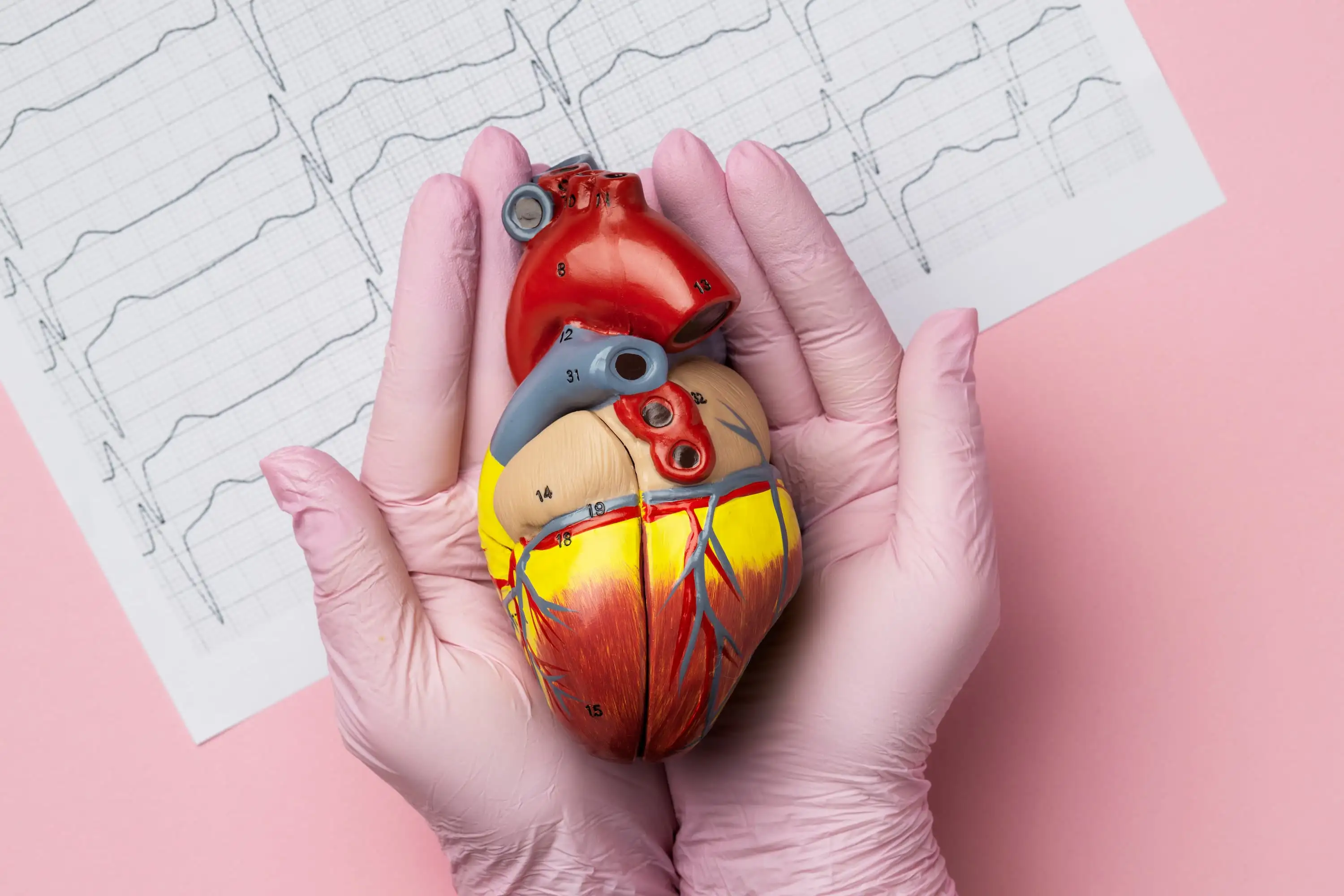
This case report highlights a rare presentation of myocardial infarction as a severe headache, known as cardiac cephalalgia, in a 47-year-old man with diabetes and tobacco use history. Despite the absence of chest pain, ECG revealed an inferior STEMI, and coronary angiography confirmed critical blockages, successfully treated with stenting. The headache resolved immediately after revascularization. The report emphasizes the importance of considering cardiac causes in people presenting with sudden, intense headaches—especially those having cardiovascular risk factors—to avoid misdiagnosis and ensure timely intervention.
Introduction
Worldwide, coronary heart disease continues to be one of the primary contributors to illness and mortality. In 2020, nearly 244.1 million people were affected by ischaemic heart disease, with men displaying a higher prevalence when compared to women. Myocardial ischaemia's classic symptoms encompass chest pain or discomfort, which may extend to the arms, jaw, or upper abdomen and can manifest either at rest or during physical activity. Nonetheless, less common signs like palpitations or even symptom-free (silent) ischaemia are also well-documented.
Cardiac cephalalgia represents a rare but notable atypical presentation of myocardial ischaemia, where the person experiences a migraine-like headache during an ischaemic episode. Given its uncommon and vague nature, especially when chest pain is absent, this ailment may be misdiagnosed or overlooked, leading to delays in appropriate care.
This case report describes a middle-aged man who presented with a headache as the only symptom of an inferior ST-segment elevation myocardial infarction, highlighting the need to consider cardiac cephalalgia as an atypical but important sign of acute coronary syndrome (ACS).
Medical History
Discussion
ACS typically manifests as left-sided, tightening chest pain radiating to the arms, neck, or jaw. However, atypical presentations like cardiac cephalalgia—headache as the primary or sole symptom—require a high level of clinical suspicion, especially in those with cardiovascular risk factors such as diabetes mellitus. This case corresponds with previously documented reports of cardiac cephalalgia. Kobata et al. described 4 people with headaches either prior to or concurrent with myocardial ischaemia.
All had common risk factors including hypertension, diabetes, hyperlipidaemia, and smoking. Similarly, a review of 30 cases by Wei and Wang found migraine-like headaches, often frontotemporal or occipital, as typical presentations. This was frequently the only symptom, especially in men over 50 with known cardiovascular risks. Headaches were accompanied by autonomic symptoms, and diagnosis was supported by ECG changes and elevated cardiac enzymes. Most cases illustrated symptom resolution following coronary intervention.
In the present case, a 47-year-old man experienced a sudden, severe headache that woke him from sleep—an uncommon presentation of ACS. Although subarachnoid haemorrhage is a primary concern in such presentations, myocardial ischaemia must also be considered in high-risk individuals after ruling out neurological causes. Non-contrast computed tomography is the preferred initial test for excluding subarachnoid haemorrhage. However, this patient’s ECG changes and haemodynamic instability confirmed STEMI, obliging urgent coronary intervention.
The abrupt onset and severity of headache ruled out post-febrile headache. Absence of neurological deficits, together with ECG findings, pointed towards cardiac cephalalgia (a rare but recognized form of myocardial ischaemia). As per the International Classification of Headache Disorders (ICHD-3), it typically presents with migraine-like headache exaggerated by exertion, occurring during ischaemia, and ameliorated by nitrates. In this case study, the patient had multiple risk factors for coronary artery disease, encompassing poorly controlled longstanding diabetes and a past history of tobacco consumption.
Diabetes is linked to exaggerated atherosclerosis and autonomic neuropathy, that may blunt typical anginal symptoms and arouse unusual manifestations like headache. Cardiac autonomic neuropathy triggers sensory denervation, which may explain why chest pain is absent in diabetic patients. Histological and imaging studies, such as m-Iodobenzylguanidine scintigraphy, have shown evidence of sympathetic denervation and altered pain perception in such individuals. Also, the lack of a circadian pattern of cardiac events in diabetics further supports the presence of autonomic dysfunction.
Differentiating cardiac cephalalgia from migraine is critical. Common migraine treatments like triptans and ergot derivatives are vasoconstrictors and contraindicated in those suffering from myocardial ischaemia. Therefore, ECG should be part of the initial assessment of any acute headache in patients with cardiovascular risk factors. Serial ECGs and cardiac biomarkers can additionally assist in identifying ischaemia. Though the exact mechanism behind cardiac cephalalgia is unclear, several hypotheses have been proposed:
In this case, the referred pain mechanism is likely, given the patient’s poorly controlled diabetes and resulting autonomic neuropathy, which likely masked chest pain and allowed headache to emerge via alternative neural pathways. An additional factor was a recent upper respiratory tract infection with throat irritation—an acknowledged risk factor for ACS, particularly within 2 weeks of infection.
The inflammatory and pro-thrombotic response may destabilize atherosclerotic plaques. Furthermore, the patient had been taking 50 mg twice daily diclofenac sodium for 2 days before admission. NSAIDs, particularly diclofenac, are associated with elevated cardiovascular risk and can trigger ACS in those with existing risk factors.
Learning
Cardiac cephalalgia is a rare but underrecognized manifestation of acute myocardial ischemia. Clinicians should include ACS in the differential diagnosis when high-risk individuals—such as those with diabetes or a smoking history—present with a sudden, intense headache even in the absence of neurological deficits or migraine history. Simple diagnostic tools like ECG and cardiac biomarkers can be lifesaving. Early recognition is key to preventing misdiagnosis and ensuring timely treatment of potentially fatal cardiac events.
BMC Cardiovascular Disorders
Cardiac cephalalgia-headache as an atypical presentation of ST-segment elevation myocardial infarction: a case report
Udayanga Andadola et al.


Comments (1)